The hunt for topotypes at Project Phoenix continues with Andrew Baird, Gus Crosbie and Hanaka Mera spending four days at the research station on Low Isles off Port Douglas on the Great Barrier Reef (GBR).
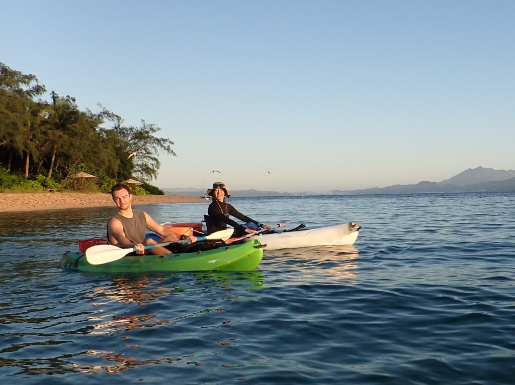
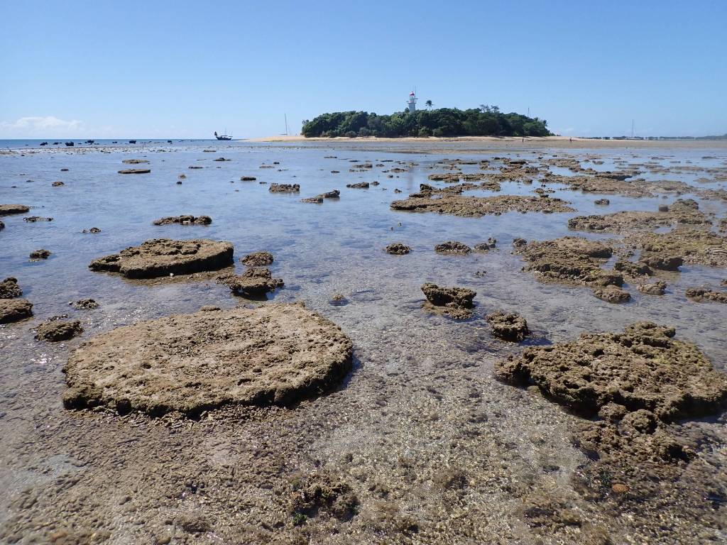
Low Isles is an important site in the history of coral reef ecology, being the site of the GBR Expedition in 1928-9 when an international group of scientists spent a year on the island doing some of the first coral reef field work ever. The group achieved much with very little equipment and in many ways not much has changed on the Island since. There are no SCUBA facilities, no boats and no aquaria, so just like the good old days, the Project Phoenix team collected by hand on snorkel from the beach or from sea kayaks. A delightful step back to a time when slow science was the norm. The research station is nestled in the shade of the forest and was comfortable and spotless clean, the reef is recovering well from recent bleaching, at least in some areas, the birds in the forest and at sea were energetic and sunsets over the Great Divide were majestic.
The team were hunting for topotypes of eight Montipora and two Porites species collected during the GBR Expedition and later named by Cyril Crossland in 1952. The hunt for topotypes is always complicated by the lack of field images taken back in the day. Therefore, when collecting one has to imagine what the colony might look like in the field based on the skeletons of the type specimens. Only once the field specimen is bleached and dried is it possible to compare it to the type and decide whether or not you have collected the same species. The example below illustrates what is often a complicated process.
One of the topotypes the team was chasing was Montipora fossae Crossland 1952. The holotype is illustrated in the figure below (Fig. 1). Crossland’s text also refers to field images in the reports of the GBR Expedition (Stephenson et al 1931) that we could also refer to (Fig. 2). Stephenson and others refer to this species as M. ramosa Bernard 1897 (Fig. 1). The skeleton and the field images suggest that M. fossae is a chunky version of what most people currently working on the GBR would call M. digitata (Dana 1846). So, a chunky M. digitata is what the team set out to look for. The closest specimen they found was numbered 74-0346 (Fig. 1).
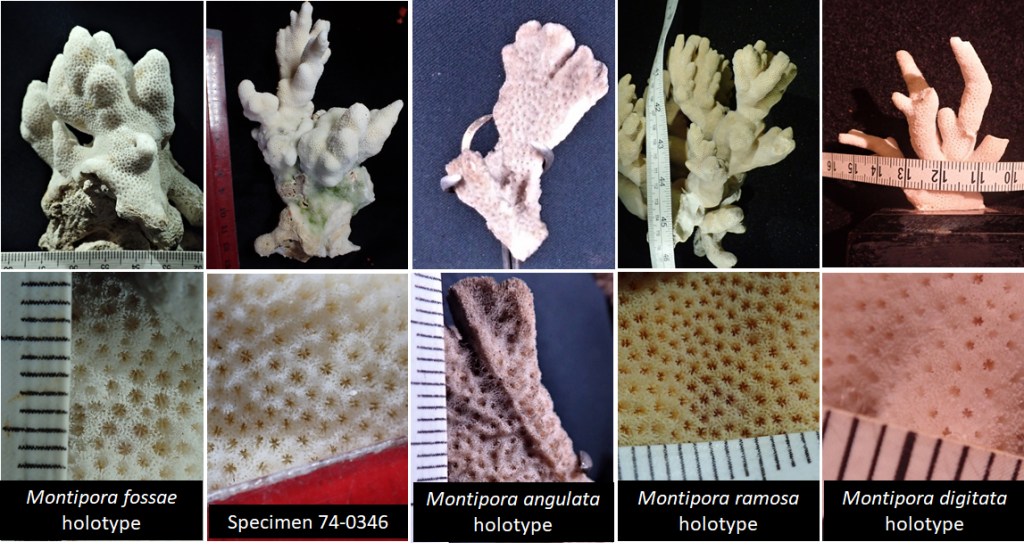

Captioned as “Montipora ramosa western moat. This looks more like Cyril’s M. fossae.”
Is this a good topotype? After a close look at the skeleton of the sample we collected and comparing to the type, the answer is probably ‘no’. The gross morphology is similar but the corallites of the holotype are flush with the surface of the coenosteum, whereas in the specimen 74-0346 they occur at the bottom of small pits (foveolate). Also, the coenosteum of the specimen 74-0346 has small elaborations, called trabeculae, whereas the coenosteum of the holotype of M. fossae is smooth, or glabrous.
What else might the field specimen 74-0346 be? Perhaps digging into taxonomic history might help.
Montipora fossae Crossland 1952 was synonymized with M. angulata (Lamark 1816; Fig. 1) by Veron & Wallace 1984. Comparing the holotypes of each suggests Veron & Wallace 1984 were in error. The branches in M. angulata are laterally compressed, whereas those of M. fossae are round; M. angulata has prominent coenosteum ridges which are absent in M. fossae; and finally the corallites are foveolate in M. angulata but not in M. fossae.
Perhaps specimen 74-0346 is M. ramosa as originally proposed by Stephenson et al (1931)? Again, the corallites are wrong as is the coenosteum. Indeed, a comparison of the types suggests that M. fossae is probably closer to M. ramosa than M. angulata.
Finally, could specimen 74-0346 be M. digitata? Highly unlikely – the corallites on the holotype of M. digitata are only half the size of those in specimen 74-0346, they are more widely spaced and the coenosteum lacks trabeculae.
In summary, the specimen is probably none of the above species and it is now necessary to compare it to the type material of the other 20 odd nominal species of small branching Montipora. And morphology is only one line of evidence for species delimitation. A molecular analysis might suggest that all these morphologies are within the range of one very plastic species.
Andrew, Gus and Hanaka thank the Low Isles caretakers Sean and Emma, the crew of the Calypso Reef Cruises and Bonnie Boyce of GBRMPA for their help in organizing the trip.
Literature cited
Bernard, H. M. 1897. The genus Montipora; the genus Anacropora. Catalogue of the Madreporarian Corals in the British Museum (Natural History) 3:1-192, pls. 191-133.
Crossland, C. 1952. Madreporaria, Hydrocorallinae, Heliopora and Tubipora. Sci. Rep. Great Barrier Reef Exped. 1928-1929 6:85-257.
Dana, J. D. 1846. Structure and classification of Zoophytes. Lea and Blanchard, Philadelphia.
Lamarck, J. B. 1816. Histoire naturelle des Animaux sans Vertèbre, Paris.
Stephenson, T. A., A. Stephenson, G. Tandy, and M. Spender. 1931. The structure and ecology of Low Isles and other reefs. Scientific Reports / Great Barrier Reef Expedition 1928-29. 3:17-112.
Veron, J. E. N., and C. C. Wallace. 1984. Scleractinia of Eastern Australia – Part V. Family Acroporidae. Australian Institute of Marine Science Monograph Series 6:1-485.
